University Notes Your Online Solution

Do not foud your proper note ? We help you with all the better Notes..

Not Found Your Notes here?

Help Us to add your Subject in University notes! Just contract with Us !

A vast of subject! Select your Catagories !

This Blank space is Only for Your Subject! So fullfill this Gap By your Note!

No Need of Paper!

Just Stay with us and Study Online.. We add all the subject day by day!!

Don't Forget Us We Stay with You!

Just bookmark our page. We are staying with U until your Final destination.

Showing posts with label Medical Science. Show all posts

Androstenedione

Androstenedione is a steroid hormone that has weak, androgenic actions on the body itself. However, it mainly acts as a stepping stone in the manufacture of testosterone and oestrogen within the body.
Alternative names for androstenedione
Andro; andros; 4-Androstenedione. 17 ketotestosterone; 4-androsten-3,17-dione.

What is androstenedione?
Androstenedione is described as a ‘pro-hormone’ because it has few effects itself.  Instead, it is important because of the ability of different parts of the body to convert it into the hormones, testosterone and oestrogen, which exert many effects on the body.

In females, the outer part of the adrenal glands (known as the cortex) and the ovaries release androstenedione into the bloodstream where it is converted to provide around half of all testosterone and almost all of the body’s oestrone, a form of oestrogen.  Although the testes produce large amounts of androstenedione in males, they secrete little of this into the blood and, instead, rapidly convert it into testosterone within the testes.  The adrenal glands also produce androstenedione in men, but this contribution is swamped by the testes’ overwhelming production of the other androgenic hormone, testosterone.   

How is androstenedione controlled?  
Due to its secretion from a number of different glands and its often rapid conversion to other hormones, the control of androstenedione within the body is very complex.  However, two key parts of the brain (the hypothalamus and pituitary gland) are known to be important in the control of androstenedione secretion from the testes, ovaries and adrenal cortex.  The release of androstenedione by the adrenal cortex is thought to be related to the pituitary gland’s secretion of a specialised hormone, adrenocorticotropic hormone.  Precisely how adrenocorticotropic hormone and other hormones control the adrenal gland’s production of androstenedione is, however, unclear.  The testes and ovaries are stimulated to release androstenedione by luteinising hormone and follicle stimulating hormone.  These are released from the anterior pituitary gland in response to a hormone signal from the hypothalamus.

What happens if I have too much androstenedione?
The effects of too much androstenedione are likely to result from its conversion in the body to oestrogen or testosterone.

In men, too much androstenedione may lead to an imbalance in oestrogen and testosterone production, leading to changes such as breast development.  Depending on the cause of the excess androstenedione, other changes, such as the testes becoming smaller, might also occur.

In women, excess body and facial hair growth (called hirsutism), stopping of periods (amenorrhoea), worsening acne and changes to the genitalia may result from too much androstenedione.

Although androstenedione is often abused by bodybuilders in an effort to build muscle bulk, a small number of studies have suggested that its long-term use may actually decrease muscle strength.  The precise consequences of having too much androstenendione are, therefore, still unclear.

Aldosterone

Alternative names for aldosterone
Electrocortin, 11β,21-dihydroxy-3,20-dioxopregn-4-en-18-al.

What is aldosterone?
Aldosterone is a hormone produced in the outer section (cortex) of the adrenal glands, which sit above the kidneys. It plays a central role in the regulation of blood pressure mainly by acting on organs such as the kidney and the colon to increase the amount of salt (sodium) reabsorbed into the bloodstream and the amount of another salt called potassium removed in the urine. Aldosterone also causes water to be reabsorbed along with sodium; this increases blood volume and therefore blood pressure. Thus, aldosterone indirectly regulates blood levels of electrolytes (sodium, potassium and hydrogen) and helps to maintain the blood pH. 

How is aldosterone controlled?  
Aldosterone is part of a group of linked hormones, which form the renin-angiotensin-aldosterone system. Activation of this system occurs when there is decrease in blood flow to the kidneys following loss of blood volume or a drop in blood pressure (eg due to a haemorrhage) or decrease in plasma sodium concentration. Renin is an enzyme that leads to a series of chemical reactions resulting in the production of angiotensin II, which in turn stimulate aldosterone release. Aldosterone causes an increase in salt and water reabsorption into the bloodstream from the kidney thereby increasing the blood volume, restoring salt levels and blood pressure. Once salt levels and blood pressure are corrected and the body becomes rehydrated, the level of renin in the bloodstream falls and therefore the amount of aldosterone in the blood also falls, meaning more water is excreted in the urine. The renin-angiotensin-aldosterone system is an example of a negative feedback system.

The other two main regulators of aldosterone secretion are increase in the plasma potassium concentration and adrenocorticotropic hormone (ACTH) secreted by the anterior pituitary, which can act via either positive or negative feedback mechanisms, depending on the extent of changes in the levels of these two regulators.

What happens if I have too much aldosterone?
The most common cause of high aldosterone levels is excess production frequently from a small benign adrenal tumour (hyperaldosteronism). The symptoms include high blood pressure, low blood levels of potassium, an abnormal increase in blood volume and sometimes the blood becomes alkaline indirectly as a consequence of aldosterone’s action in promoting acid secretion.

What happens if I have too little aldosterone?
There are two conditions where there are low aldosterone levels.

In Addison's disease, there is a general loss of adrenal function resulting in low blood pressure, lethargy and an increase in potassium levels in the blood (see the article on Addison's disease for further information).

An enzyme called aldosterone synthase is responsible for the last steps in the production of aldosterone. Rare mutations in the gene that codes for aldosterone synthase can result in low or absent production of aldosterone (aldosterone synthase deficiency). With this rare genetic condition, the symptoms are similar to that of Addison's disease but milder

ACTH

Alternative names for adrenocorticotropic hormone
ACTH; adrenocorticotrophin; corticotropin.

What is adrenocorticotropic hormone?
Adrenocorticotropic hormone is made in the corticotroph cells of the anterior pituitary gland.  It is secreted in several intermittent pulses during the day into the bloodstream and transported around the body.  Like cortisol, levels of adrenocorticotropic hormone are generally high in the morning when we wake up and fall throughout the day.  This is called a diurnal rhythm.  Once adrenocorticotropic hormone reaches the adrenal glands, it binds on to receptors causing the adrenal glands to secrete more cortisol, resulting in higher levels of cortisol in the blood.  It also increases production of the chemical compounds that trigger an increase in other hormones such as adrenaline and noradrenaline.

How is adrenocorticotropic hormone controlled?
Secretion of adrenocorticotropic hormone is controlled by three inter-communicating regions of the body, the hypothalamus, the pituitary gland and the adrenal glands. This is called the hypothalamic-pituitary-adrenal axis.  When adrenocorticotropic hormone levels in the blood are low, a group of cells in the hypothalamus release a hormone called corticotrophin-releasing hormone which stimulates the pituitary gland to secrete adrenocorticotropic hormone into the bloodstream.  High levels of adrenocorticotropic hormone are detected by the adrenal glands which stimulate the secretion of cortisol, causing blood levels of cortisol to rise.  As the cortisol levels rise, they start to slow down the release of corticotrophin-releasing hormone from the hypothalamus and adrenocorticotropic hormone from the pituitary gland.  As a result, the adrenocorticotropic hormone levels start to fall.  This is called a negative feedback loop.

Stress, both physical and psychological, also stimulates adrenocorticotropic hormone production and hence increases cortisol levels.

What happens if I have too much adrenocorticotropic hormone?
The effects of too much adrenocorticotropic hormone are mainly due to the increase in cortisol levels which result.  Higher than normal levels of adrenocorticotropic hormone may be due to:

Cushing's disease – this is the most common cause of increased adrenocorticotropic hormone. It is caused by a non-cancerous tumour called an adenoma located in the pituitary gland which produces excess amounts of adrenocorticotropic hormone. (Please note, Cushing’s disease is just one of the numerous causes of Cushing’s syndrome).
 
A tumour, outside the pituitary gland, producing adrenocorticotropic hormone (also called ectopic adrenocorticotropic hormone tumour).
 
Addison's disease (although cortisol levels are low, adrenocorticotropic hormone levels are raised).
 
Congenital adrenal hyperplasia (a genetic disorder with inadequate production of cortisol, aldosterone or both).
What happens if I have too little adrenocorticotropic hormone?
Lower than normal levels of adrenocorticotropic hormone may be due to:

Cushing's syndrome related to an adrenal tumour
 
Cushing's syndrome due to steroid medication
 
Conditions affecting the pituitary gland, eg, hypopituitarism
 
Side-effect of pituitary surgery or radiation therapy
Too little adrenocorticotropic hormone could lead to a poorly functioning adrenal gland or even Addison’s disease, due to insufficient production of cortisol.  Symptoms may include fatigue, dizziness (especially upon standing), weight loss, muscle weakness, mood changes and darkened areas of skin.

Adrenaline hormone

Alternative names for adrenaline
Epinephrine.

What is adrenaline?
Adrenaline and noradrenline are two separate but related hormones and neurotransmitters. They are produced in the medulla of the adrenal glands and in some neurons of the central nervous system. They are released into the bloodstream and serve as chemical mediators, and also convey the nerve impulses to various organs. Adrenaline has many different actions depending on the type of cells it is acting upon.  However, the overall effect of adrenaline is to prepare the body for the ‘fight or flight’ response in times of stress, ie, for vigorous and/or sudden action. Key actions of adrenaline include increasing the heart rate, increasing blood pressure, expanding the air passages of the lungs, enlarging the pupil in the eye (see figure), redistributing blood to the muscles and altering the body’s metabolism, so as to maximise blood glucose levels (primarily for the brain). A closely related hormone, noradrenaline, is released mainly from the nerve endings of the sympathetic nervous system (as well as in relatively small amounts from the adrenal medulla). There is a continuous low level of activity of the sympathetic nervous system resulting in release of noradrenaline into the circulation, but adrenaline release is only increased at times of acute stress.

Adrenaline, like noradrenaline, is also used as a transmitter by some nerve cells to communicate with other cells (a neurotransmitter). Very little adrenaline is used in this way.

How is adrenaline controlled?
Adrenaline is mainly released in response to stressful events to prepare the body for the ‘fight or flight’ response. These events lead to the activation of nerves connected to the adrenal glands, which trigger the secretion of adrenaline and thus increase the levels of adrenaline in the blood. This process happens relatively quickly, within 2 to 3 minutes of the stressful event being encountered. When the stressful situation ends, the nerve impulses to the adrenal glands are lowered, meaning that the adrenal glands stop producing adrenaline.

Stress also stimulates the release of adrenocorticotropic hormone from the pituitary gland, which promotes the production of the steroid hormone cortisol from the cortex of the adrenal glands. This steroid hormone is more important in altering the body’s metabolism (ie, raising plasma glucose) under conditions of longer-term, ongoing (chronic), rather than acute, stress.

What happens if I have too much adrenaline?
Overproduction of adrenaline is rare. Too much adrenaline can be caused by a variety of things, including a rare tumour of the adrenal medulla (a phaeochromocytoma). Symptoms may include rapid heart beat, high blood pressure, anxiety, weight loss, excessive sweating and palpitations.

What happens if I have too little adrenaline?
Suffering from too little adrenaline is very unusual. Among other things, it would result in an inability to prepare the body for action in response to a stressful or physically demanding situation.

Short Note : Oxytocin

Alternative names for oxytocin
Alpha-hypophamine; manufactured versions – carbetocin, syntocinon and pitocin.

What is oxytocin?
Oxytocin is produced in the hypothalamus and is secreted into the bloodstream by the posterior pituitary
gland. Secretion depends on electrical activity of neurons in the hypothalamus – it is released into the
blood when these cells are excited.
The two main actions of oxytocin in the body are contraction of the womb (uterus) during childbirth and
lactation. Oxytocin stimulates the uterine muscles to contract and also increases production of
prostaglandins which increase the contractions further. Manufactured oxytocin is sometimes given to
induce labour if it has not started naturally or it can be used to strengthen contractions to aid childbirth.
In addition, manufactured oxytocin is often given to speed up delivery of the placenta and reduce the risk
of heavy bleeding by contracting the uterus. During breastfeeding, oxytocin promotes the movement of
milk into the breast, allowing it to be excreted by the nipple. Oxytocin is also present in men, playing a
role in sperm movement and production of testosterone by the testes.
More recently, oxytocin has been suggested to be an important player in social behaviour.
In the brain, oxytocin acts as a chemical messenger and has been shown to be important in human
behaviours including sexual arousal, recognition, trust, anxiety and mother-infant bonding. As a result,
oxytocin has been called the ‘love hormone’ or ‘cuddle chemical’.
Many research projects are undertaken, looking at the role of oxytocin in addiction, brain injury, anorexia
and stress amongst other topics.

How is oxytocin controlled?
Oxytocin is controlled by a positive feedback mechanism where release of the hormone causes an action
which stimulates more of its own release. When contraction of the uterus starts, for example, oxytocin is
released which stimulates more contractions and more oxytocin to be released. In this way, contractions
increase in intensity and frequency.
There is also a positive feedback involved in the milk-ejection reflex. When a baby sucks at the breast of
its mother, the stimulation leads to oxytocin secretion into the blood which then causes milk to be let
down into the breast. Oxytocin is also released into the brain to help stimulate further oxytocin
secretion. These processes are self-limiting; production of the hormone is stopped after the baby is
delivered or when the baby stops feeding.

What happens if I have too much oxytocin?
At present, the implications of having too much oxytocin are not clear. High levels have been linked to
benign prostatic hyperplasia, a condition which affects the prostate in more than half of men over the age
of 50. This may cause difficulty in passing urine.
It may be possible to treat this condition by manipulating oxytocin levels; however, more research is
needed before any possible treatments are available.

What happens if I have too little oxytocin?
Similarly, it is not fully understood at present if there are any implications of having too little oxytocin in
the body. A lack of oxytocin in a nursing mother would prevent the milk-ejection reflex and prevent
breastfeeding.
Low oxytocin levels have been linked to autism and autistic spectrum disorders (eg, Asperger syndrome) –
a key element of these disorders being poor social functioning. Some scientists believe oxytocin could be
used to treat these disorders. In addition, low oxytocin has been linked to depressive symptoms and it has
been proposed as a treatment for depressive disorders. However, there is not enough evidence at present
to support its use for any of these conditions.

10 Chemical Element of Human Body


Here's a look at the chemical composition of the human body, including element abundance and how each element is used. Elements are listed in order of decreasing abundance, with the most common element (by mass) listed first. Approximately 96% of body weight consists of only four elements: oxygen, carbon, hydrogen, and nitrogen. Calcium, phosphorus, magnesium, sodium, potassium, chlorine, and sulfur are macronutrients or elements the body needs in a significant amount.

Crystals of elemental magnesium. - Warut Roonguthai


1.  Oxygen

By mass, oxygen is the most abundant element in the human body. If you think about it, this makes sense, since most of the body consists of water or H2O. Oxygen accounts for 61-65% of the mass of the human body. Even though there are many more atoms of hydrogen in your body than oxygen, each oxygen atom is 16 times more massive than a hydrogen atom.

2.  Carbon

All living organisms contain carbon, which forms the basis for all of the organic molecules in the body. Carbon is the second most abundant element in the human body, accounting for 18% of body weight.


3.  Hydrogen

Hydrogen accounts for 10% of the mass of the human body.

4.  Nitrogen

Approximately 3% of the mass of the human body is nitrogen.

5.  Calcium

Calcium accounts for 1.5% of human body weight.

6.  Phosphorus

About 1.2% to 1.5% of your body consists of phosphorus.

7.  Potassium

Potassium makes up 0.2% to 0.35% of the adult human body.

8.  Sulfur

Sulfur's abundance is 0.20% to 0.25% in the human body.

9.  Sodium

Approximately 0.10% to 0.15% of your body mass is the element sodium.

10.  Magnesium

The metal magnesium comprises about 0.05% of human body weight.
 

Rapid Diagnostic Technique of Virus

RAPID DIAGNOSTIC TECHNIQUES
These include:
1. Electron microscopy
2. Viral nucleic acid detection
3. Serology/detection of viral antigen and antibody
1. Electron microscopy
This is used to demonstrate the presence of virus in clinical specimens and to study the
morphology or symmetry of the virus. With electron microscopy, it is possible to
recognized mixed viral infection. It is also used for detection of non-viable viruses and
those that cannot be grown in vitro. However, equipment required for the procedure is
rather expensive. Besides, large number of viral particles in excess of 106/ml must be
present in the sample before they can be detected. It is difficult to differentiate viruses
with similar morphology especially those from the same family with electron
microscopy.
Method:
Homogenize sample.
Centrifuge at low speed to remove large particulate debris.
Ultracentrifugation to sediment available virus particles.
Negative staining with heavy metal compound such as phosphotungstic acid or uranyl
acetate. (negative staining stains the background to increase contrast so that bright
virion stand out against a dark background).
Addition of immune serum (immunoelectron microscopy) to increase sensitivity by
clumping/agglutinating virus particles and enhance recovery following centrifugation).
Observe at X13,000-100,000 magnification.
2. Detection of viral nucleic acid:
Oligonucleotide probes are used for the detection of viral DNA or RNA. Insufficient viral
nucleic acid in sample is increased by amplification using polymerase chain reaction.
Probes anneal to the targeted viral nucleic acid sequence which can be the whole genome,
specific gene or nucleic acid segment. Variable or conserved sequence can be targeted.
Double stranded genomes are first separated by heating. Oligonucleotide probes are
labeled with radioactive isotopes such as 32P or 35S to allow viewing. Non-radioactive
labels such as alkaline phosphatase fluorescein and horse radish peroxidase can be used
for direct viewing whilebiotin and digoxigenin are used for indirect viewing.
I. Dot-blot hybridization
Nucleic acid, usually DNA is extracted from sample
Extracted nucleic acid is spotted directly onto charged nylon or nitrocellulose membrane
Nucleic acid binds firmly onto membrane after baking
Fluorescent dye- or radioisotope- labeled probe is added
The membrane is washed to remove unbound materials
Binding of probe to targeted nucleic acid is detected by autoradiography or by colour
precipitation
II. In-situ hybridization
Viral nucleic as is detected in frozen section of infected cells with the aid of labeled
oligonucleotide probes. Intracellular location of viral nucleic acid is revealed by
autoradiography or immunoperoxidase cytochemistry
III. Southern blot hybridization
Restriction enzymes are used to cleave DNA into short oligonucleotides
Oligonucleotides are separated by agarose electrophoresis or acrylamide gel
electrophoresis
Separated oligonucleotides are transferred by blotting onto nitrocellulose
membrane or nylon
Probes are added and reaction detected by autoradiography or by colour
development
IV. Northern blot hybridization
RNA hybridization similar to southern blot
V. Western blot
Application to protein identification
VI. Polymerase chain reaction
Extraction of DNA or RNA nucleic acid from sample
Amplification of the extracted nucleic acid in a series of repeated cycles of denaturation,
primer annealing and polymerization using heat-resistant Taq (Thermus aquaticus)
polymerase. Specific primers recognize and bind to the targeted gene to initiate the
amplification reaction.
Amplified sequences are stained with ethidium bromide and separated by
electrophoresis
The separated sequence is viewed under ultraviolet transillumination.

This procedure is very specific and highly sensitive

Diagnosis of Viral Diseases

DIAGNOSIS OF VIRAL DISEASESMicrobiology এর চিত্র ফলাফল
There are well over 1000 known viruses of vertebrate and it is impossible for a single
laboratory to have all the resources required for the diagnosis of all these viruses.
Laboratories tend to specialize on particular viruses and serve as reference laboratories.
Stages of viral disease diagnosis:
1. Clinic: presumptive diagnosis based on clinical signs and history
2. Pathology: Observed lesions and pathological changes at gross and histopathological
levels. History may provide clues.
3. Microbiological diagnosis: confirmatory diagnosis
Principles of microbiological diagnosis:
i. Isolation of viruses
ii. Detection of viral nucleic acid/specific genes
iii. Detection of viral antigen
iv. Detection of specific virus-induced antibody
Sample collection:
a. Samples must be collected at from the right site and at the right time
b. Samples to be collected must relate to the clinical signs and pathological changes
observed
44
c. Samples must be collected as soon as clinical signs are observed
d. Knowledge of the pathogenesis of the disease may dictate the type of sample to be
collected
e. Proper labeling of samples for identification and to avoid confusion
f. Samples must be sent to the laboratory with history and tentative diagnosis
g. Transport samples with ice packs (4 oC) if transit time is less than 24 hours
h. For transit above 24 hours, use dry ice at -70 OC.
i. Long term storage is achieved with liquid nitrogen at -196 OC.
j. Use transport medium containing buffer (isotonic saline) with bovine albumin/foetal calf
serum (protein to prolong virus survival), antibiotic and antifungal agent (to prevent
contaminants)
Samples for diagnosis of viral diseases:
Respiratory tract infection: nasal swab, tracheal swab, nasopharyngeal aspirate, lung
tissue.
Enteric infection: Faeces, rectal swab.
Genital tract infection: prepucial washing, semen, genital swab.
Eye infection: Conjunctival swab.
Skin infection: Vesicular fluid, epithelial scrapings, biopsy of solid lesions.
Central nervous system: cerebrospinal fluid, faeces, nasal swab, brain tissue.
Generalized infection: Nasal swab, faeces, blood leukocytes.
Post mortem examination: relevant organ.

Every case: Blood for serum to be used in serology.

Classification of Viruses

CLASSIFICATION OF VIRUSES
Following the discovery of viruses, earliest studies on them were based on their
filterability, and observations on the diseases which they caused. Early classification
systems were premised on pathogenic effects of the viruses and their transmission
patterns. However, with the invention of electron microscope and sophisticated molecular
techniques that permitted ultra-structural studies, details of the structures and
compositions of viruses began to emerge. Thereafter, it became possible to group viruses
on the basis of shared features of the virions.
Consequently, the following general parameters are have been used for classification of
viruses:
1. Pathogenicity
2. Ecological characteristics
3. Physico-chemical characteristics

Pathogenicity:

In this classification, viruses affecting same tissues producing similar syndrome and
pathological manifestations are grouped together.
- Viruses affecting the respiratory tract: Influenza, rhinoviruses, parainfluenza, adenovirus
- Vesicular viruses: Foot-and-Mouth-Disease, vesicular stomatitis
- Central nervous system viruses: rabies, equine encephalitis, entroviruses (polio), mumps
- Mucous membrane viruses: Myxomaviruses
- Enteric viruses: rotaviruses,
- Limitations of this classification system: some viruses affect more than one system of
the body and they will belong to several groups. Pantropic viruses affecting multiple
systems such as canine distemper, Newcastle disease, rinderpest, pestes des petits
ruminants will belong to respiratory, enteric and CNS groups.

Ecological characteristics:

Ecological features of viruses such as the involvement of vectors or vertebrate reservoirs
in their transmission cycles and maintenance in nature can be used for classification.
Viruses are classified into arboviruses and non-arboviruses or roboviruses and nonroboviruses.
Arboviruses: these are viruses that are transmitted biologically between blood sucking
arthropods (such as ticks, culicoides, mosquitoes) and vertebrate hosts. They cause
disease in the vertebrate host but not in the arthropods. Examples include African swine
36
fever virus (soft ticks), Yellow fever virus (mosquitoes), Equine encephalitis virus
(mosquitoes), African horse sickness virus (culicoides).
Roboviruses: These are viruses with rodent reservoirs. Infected rodents are
asymptomatic. They shed the virus in their urine and contaminate the human
surroundings, food, drinks and formites. Example include Lassa fever virus with rat as

reservoir.
Microbiology এর চিত্র ফলাফল

Physico-chemical characteristics:
These are the most reliable, verifiable and satisfactory parameters for classifying viruses.
Viruses are classified based on the following criteria:
Type of nucleic acid (RNA or DNA)
Number of strands of the nucleic acid (single or double stranded)
Physical construction of the nucleic acid (linear, circular, circular with break, segmented,
non-segmented)
Polarity of the viral genome: positive polarity (viral genome can be used directly as
mRNA) and negative polarity(viral genome must be transcribed into mRNA)
Symmetry of the nucleocapsid
Presence or absence of envelope
Size of the virus
Antigenic and chemical compositions
Susceptibility to physical and chemical changes
Based on these criteria, viruses are grouped into families, subfamilies and genera. Further
subdivision is based on the degree of antigenic similarity and serological tests

Diagnosis of Fungi infection

Microbiology এর চিত্র ফলাফল
1. Skin scrapings suspected to contain dermatophytes or pus from a lesion can be
mounted in KOH on a slide and examined directly under a microscope.
2. Skin testing (dermal hypersentivity) used to be popular as a diagnostic tool, but
this use is now discouraged because skin test may interfere with serological
studies by causing false positive results. It may still be used to evaluate the
patient’s immunity as well as a population exposure index in epidemiological
studies.
3. Serology may be helpful when it is applied to a specific fungal disease: there are
no screening antigens for “fungi” in general. Because fungi are poor antigen, the
efficacy of serology varies with different fungal infections. The serologic test will
be discussed under each mycosis. The most common serological tests for fungi
are based on latex agglutination, double immunodiffusion, complement fixation
and enzyme immunoassays. While latex agglutination may favor the detection of
IgM antibodies, double immunodiffusion and complement fixation usually detect
IgG antibodies. Some EIA tests are being developed to detect both IgG and IgM
antibodies. There are some tests which can detect specific fungal antigens, but
they are just coming into general use.
4. Direct fluorescent microscopy may be used for identification, even on non-viable
cultures or on fixed tissue secton. The reagents for this test are difficult to obtain.
5. Biopsy and histopathology. A biopsy may be very useful for the identification and
as a source of the tissue-invading fungi. Usually the Gomori methenamine silver
(GMS) stain is used to reveal the organism which stain black against a green
background. The H&E stain does not always tint the organism, but it will stain the
inflammatory cells.
6. Culture. A definite diagnosis requires a culture and identification. Pathogenic
fungi are usually grown on Saboouraud dextrose agar. It has a slightly acidic pH
(5,6), cyclohezamide, penicillin, streptomycin or other inhibitory antibiotic are
often added to prevent bacterial contamination and overgrowth. Two cultures are
25
inoculated and incubated separately at 250C and 370C to reveal dimorphism. The
cultures are examined macroscopically and microscopically. They are not
considered negative for growth until ahter 4 weeks of incubation.
Reference:
Betsy, T and Keogh, J: (2005) Microbiology demystified. Published by the McGraw-Hill

Companies, New York, USA.

Digestive System Glossary of Human

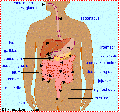
digestive system
abdomen - the part of the body that contains the digestive organs. In human beings, this is between the diaphragm and the pelvis
alimentary canal - the passage through which food passes, including the mouth, esophagus, stomach, intestines, and anus.
anus - the opening at the end of the digestive system from which feces (waste) exits the body.
appendix - a small sac located on the cecum.
ascending colon - the part of the large intestine that run upwards; it is located after the cecum.
bile - a digestive chemical that is produced in the liver, stored in the gall bladder, and secreted into the small intestine.
cecum - the first part of the large intestine; the appendix is connected to the cecum.
chyme - food in the stomach that is partly digested and mixed with stomach acids. Chyme goes on to the small intestine for further digestion.
descending colon - the part of the large intestine that run downwards after the transverse colon and before the sigmoid colon.
digestive system - (also called the gastrointestinal tract or GI tract) the system of the body that processes food and gets rid of waste.
duodenum - the first part of the small intestine; it is C-shaped and runs from the stomach to the jejunum.
epiglottis - the flap at the back of the tongue that keeps chewed food from going down the windpipe to the lungs. When you swallow, the epiglottis automatically closes. When you breathe, the epiglottis opens so that air can go in and out of the windpipe.
esophagus - the long tube between the mouth and the stomach. It uses rhythmic muscle movements (called peristalsis) to force food from the throat into the stomach.
gall bladder - a small, sac-like organ located by the duodenum. It stores and releases bile (a digestive chemical which is produced in the liver) into the small intestine.
gastrointestinal tract - (also called the GI tract or digestive system) the system of the body that processes food and gets rid of waste.
ileum - the last part of the small intestine before the large intestine begins.
intestines - the part of the alimentary canal located between the stomach and the anus.
jejunum - the long, coiled mid-section of the small intestine; it is between the duodenum and the ileum.
liver - a large organ located above and in front of the stomach. It filters toxins from the blood, and makes bile (which breaks down fats) and some blood proteins.
mouth - the first part of the digestive system, where food enters the body. Chewing and salivary enzymes in the mouth are the beginning of the digestive process (breaking down the food).
pancreas - an enzyme-producing gland located below the stomach and above the intestines. Enzymes from the pancreas help in the digestion of carbohydrates, fats and proteins in the small intestine.
peristalsis - rhythmic muscle movements that force food in the esophagus from the throat into the stomach. Peristalsis is involuntary - you cannot control it. It is also what allows you to eat and drink while upside-down.
rectum - the lower part of the large intestine, where feces are stored before they are excreted.
salivary glands - glands located in the mouth that produce saliva. Saliva contains enzymes that break down carbohydrates (starch) into smaller molecules.
sigmoid colon - the part of the large intestine between the descending colon and the rectum.
stomach - a sack-like, muscular organ that is attached to the esophagus. Both chemical and mechanical digestion takes place in the stomach. When food enters the stomach, it is churned in a bath of acids and enzymes.
transverse colon - the part of the large intestine that runs horizontally across the abdomen.

History of pathology


- The oldest civilized people (Chinese, Indians, Egyptians) were under the impression that disease occurred when demons or evil spirits were displeased with an individual. The medicine men were concerned with appeasing these evil spirits.

 -- During the Tigris-Euphrates era, veterinary medicine was first mentioned in recorded history. Veterinary medicine received a considerable degree of attention because of the importance of the horse to society. The "Laws of Hammurabi" (2100 B.C.) established rigid rules and regulations for veterinary practitioners, and these rules included the regulation of fees. In addition, the Hebrews, Babylonians, Greeks and Carthaginians made important contributions to the medical literature. However, the majority of this recorded history was destroyed by the Crusades and barbarians.

 -- The Egyptians began to influence medicine around 4000 B.C. These people were adept in certain phases of medicine, especially skull surgery. In addition,
embalming was an art employed by the Egyptians; many of these bodies (mummies) are still well preserved. There is no record of the Egyptians finding lesions or diseases during the embalming process.

 -- The Mosaic Doctrine (1500 B.C.) is the first recorded evidence of systematic meat inspection. These lays, as formulated by Moses and described in the books of Exodus and Leviticus, are similar to those we have today. Even though the Hebrews were advanced in food sanitation, they still accepted "divine displeasure" as the cause of disease.

 -- The Greek culture had a profound effect on the scientific approach to medicine. Greek physicians elucidated the principles of exact and careful clinical observations. However, they did not deal with the nature or the changes that occurred subsequent to disease.

1. HIPPOCRATES (460-375 B.C.)
AGreek physician, introduced the humoral theory of disease. He recognized four humors or fluids in the body:
 (1) blood which came from the heart;
 (2) phlegm which came from the brain;
 (3) yellow bile which came from the liver; and
 (4) black bile which came from the spleen.
Health was thought to be due to proper mixing of these four humors, while disease resulted from improper mixing. The humoral theory of disease was supported by three critical observations:
 (1) diseases were often characterized by increased discharge of fluid (perspiration, fever, vomition, diarrhea, catarrhal discharge, exudation, and transudation);
 (2) blood was the vital tissue and the individual died if exsanquinated;
 (3) the coagulation of blood was different in healthy and sick individuals.
Also, what was called phlegm is the same as fibrin. Those factors considered to be the effects of disease today were considered as causes by the humoral pathologists. The early humoral pathologists were not permitted to perform postmortem examinations on humans; thus, a confused concept of normal anatomy existed. Hippocrates is considered to be the Father of Medicine.

2. ARISTOTLE (384-323 B.C.)
A Greek philosopher, was the originator of modern anatomy and physiology. Also, he is considered to be the Father of Zoology. Aristotle dissected many animals, carried out experiments in physiology, and studied the growth and development of animal life (human autopsy examinations were forbidden during this period).

3. CLAUDIUS GALEN (129-201 A.D.)
A Greek physician practicing in Rome, was a follower of the beliefs of Hippocrates. Humoral pathology was brought to its height and most extreme development by Galen. Also, he wrote numerous medical documents and held despotic authority over European medicine for thirteen centuries after his death. Also, Galen is remembered for his views on meat inspection. He insisted that animals used for human food should be inspected prior to slaughter.

4. CORNELIUS CELSUS (30 B.C.-38 A.D.)
Was not a physician, but a man of leisure with a variety of interests. A great deal of the history of the early humoral pathologists was recorded in his work. A great number of the conditions recognized today are described in the work of Celsus. His writings described and discussed the cardinal signs of inflammation (redness, swelling, heat and pain).

5. RENATUS VEGETIUS (450-500 A.D.)
A Roman veterinarian, is credited with being the first author to write a textbook devoted exclusively to veterinary medicine. He was among the first to urge people to disregard Divine Displeasure as the cause of disease and to base their treatment and concepts of disease on a thorough knowledge of anatomy, surgery and medicine. Vegetius is considered to be the Father of Veterinary Medicine.
 -- During the middle ages, medicine entered into an era of nonproductivity and few contributions were made. However, the Renaissance brought forth profound advances in medicine and pathology. The works of Galen were questioned and new investigations were made. During this period, Divine Displeasure as the cause of disease began to gradually disappear.

6. WILLIAM HARVEY (1578-1657)
Described the blood vascular system and the circulation of blood in 1628. His works have had a far reaching effect on medicine and pathology.

7. ANTONY VAN LEEUWENHOEK (1632-1723)
Was the first to show that the microscope had practical importance in the study of tissues and other small objects (he is not credited with discovering the microscope).

8. JEAN FERNEL (1497-1558)
A Frenchman, was one of the first to describe diseases according to organs or parts of the body. he generally divided his diseases into those affecting parts above the
diaphragm, those involving parts below the diaphragm and external diseases. One of his books, entitled Pathologiae Libri, was the first medical work to be called a text of pathology.

9. GIOVANNI MORGAGNI (1682-1771)
An Italian, is recognized as one of the earliest pathologists and the originator of modern pathology. He was the first to correlate pathologic changes in the dead individual with clinical signs and symptoms shown by the individual during life.

10. MARIE-FRANCOIS XAVIER BICHAT (1771-1801)
A Frenchman, is credited with establishing the foundation for the study of histology, even though most of his work was done by physical and chemical methods (he did not possess a microscope). Bichat presented a new concept of anatomy and showed that the body was composed of twenty-one (21) tissues (vascular, osseous, muscular, cartilaginous, etc.). He is considered to be the Father of Histology.
 -- Modern veterinary medicine originated in France and the first modern veterinary school was established in Lyon, France on January 1, 1762.

11. JACQUES LABRESSIE DE SOLLEYSEL (1617-1680)
Published the first complete veterinary classic of this period, entitled Le Parfait Marechal. In this publication, Solleysel pointed out the adverse situation created by allowing the veterinary art to fall so completely into the hand of the Farrier. This book marks the beginning of the end of the horseshoer's regimen and control of veterinary medicine.

12. CLAUDE BOURGELAT (1712-1779)
A French veterinarian, was gifted in equine husbandry and wrote a book, entitled Elements of Hippiartry and the New Knowledge of Equine Medicine. He investigated and was successful in eradicating an outbreak of glanders in French Calvary horses. He was instrumental in establishing the first modern veterinary school in Lyon, France. In addition, Bourgelat established the veterinary school known as L-Ecole Veterinaire Nationale d'Alfort, located near Paris, France.

13. CARL ROKITANSKY (1804-1878)
A German, is considered as the supreme descriptive pathologist of all time. He firmly established the structural basis of disease as well as necropsy technique. However, he explained practically all diseases on the basis of blood anomalies.
 -- The field of pathology was completely reformed by cellular teachings which originated in Germany. The inspiration for this development came from Johannes
 
Mueller (1801-1858) who had many famous students (Schwann, Henle, Virchow and Schleiden).

14. RUDOLPH VIRCHOW (1821-1902)
Is known as the Father of Cellular Pathology.
He coined and explained many of the terms and concepts used today in pathology (amyloidosis, fatty degeneration, etc.). In addition, he started publication of "Virchow's Archives," a journal that has been in continuous publication since 1847. This is considered to be one of the most complete works of pathology in existence.

15. LOUIS PASTEUR
Frenchman, was one of the originators of the field of bacteriology. He demonstrated the importance of infectious organisms (bacterial) in disease. Pasteur studied human and animal diseases (pasteurellosis, anthrax, rabies, etc.) and showed that individuals could be successfully immunized by vaccines prepared from organisms.

16. ROBERT KOCH (1843-1910)
A German bacteriologist, established the Koch's Postulate, a procedure employed for proving a specific microorganism as the cause of a disease. He was the first to use artificial solid media in the attainment of pure cultures.

17. EDWIN KLEBS (1834-1913)
A student of Virchow, demonstrated the importance of bacteria in pathology.

18. JULIUS COHNHEIM (1839-1884)
A student of Virchow, is credited with being the originator of modern experimental pathology. He revealed the vascular alterations that are the basis of the inflammatory response.

19. WILLIAM H. WELCH (1850-1934)
A student of Cohnheim, is credited with bringing pathology to the United States. He was Professor of Pathology at John Hopkins University in Baltimore, Maryland.


REFERENCE: Principles of Veterinary Pathology; Runnels, Monlux and Monlux, 7th Edition, Chapter 2, pp. 6-21.

Kidney Worm : Dioctophyma

Genus: Dioctophyma  (kidney worm)
Sp: D.  renale.
Location: Kidney parenchyma.
Final host: Dog , fox, mink occasionally horse ,cattle, man.
I/H: Aquatic annelids.
 
Morphology: largest nematode of domestic animals.(103 cm.)
Females > 60 cm.diameter 1cm.
Worms has blood red colour.
Eggs are barrel shaped & brownish –yellow.
Life cycle: Adult in the kidney---egg in urine in chain / clumps—egg ingested by annelid I/H.---—L-1=L-2=L-3(with in annelid)---I/H is swallowed by dog, fox –
                                                                                                                     Eaten by
                           Annelid with L-3 taken by frog, fish---        Para tonic host
                              
                                                              L-3 penetrate bowel wall---enter body cavity—                                                                                                                                                                                                                                                                                                                                                                                                   penetrate kidney.

Pathology:
Destruction of kidney parenchyma , leaving only the capsule as a sac containing the worms.
Usually no clinical sign as normal kidney serve the function.
Kidney trouble together with nervous sign.
Retention of urine & death from uremia when bladder or urethra is bloked.
In man ,renal colic , pyuria, haematuria may occure.
Right kidney are more frequently invaded.

Diagnosis: Egg in urine.

Treatment: Surgical removal of worms.


Eye worm : Thelazia

*Genus: Thelazia  (Eye worm )
Sp:   T.  rhodeshi—Cattle, sheep, goat, buffalo.
                                  T .  lacrymalis---Horse
         T.   californiensis---Sheep, goat, cat, man
I/H: Musca sp.
      Fannia  sp.
     Morellia  sp.

Morphology: Small, thin, white worm, 1-2 c.m. long. Cuticle has prominent transverse striation at the anterior end. Females are viviparous. (Not ova-viviparous)
Location: Congunctival sac & lacrimal duct & under nictitating membrane.
Life cycle:
 Adult female in eye. --- lay L-1 in lacrimal secretion----Ingested by I/H
( files) feeding on secretion ----L3 develop in the ovarian follicle of fly.----L-migrate to proboscis--- deposited in the eye of new host.---L-4 ----Adult.
P.P.P.= 3-11 weeks.


Pathogenesis:
        Due to secreted cuticle of the worm & for the movement by active young adult.
     Lacrimation followed by conjunctivitis   Keratitis, cloudiness & ulceration of cornea.
     Swollen eye covered with exudates & pus .* Cranial opacity.
     Congestion of  conjunctiva & cornea.  * Photophobia.
     Files are usually clustered around the eye because of excessive secretion.
Diagnosis:
Observation of parasite in conjunctival sac
Presence of L-1 in lacrimal secretion.
Adult parasite in the eye can be found it manipulated by using local anaesthetic.
 
Treatment:
        Manual  removal of parasite with a fine forceps using local anaesthetic.
Anthelmintic drugs :
1.      Methyridine –20 ml s/c inj.
2.      Tetramisole—15 mg /kg body weight       orally or parent rally
3.      ,evamisole--% mg/ kg body weight
or, 1% aqueous solution –topical preparation.


Prevention:   Difficult as fly vectors are ubiquitous.

google.com